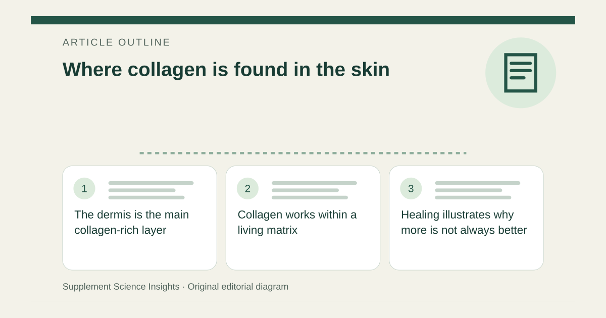
The dermis is the main collagen-rich layer
The outer skin layer is the epidermis. Beneath it lies the dermis, a connective-tissue layer that gives the skin strength and structural support. NIH's educational overview identifies collagen as a major component of this dermal framework. [1]
Below the dermis is subcutaneous tissue containing substantial fat. These layers have different functions and composition. A skin diagram should therefore place the main collagen network beneath the epidermis rather than depict it as a loose coating on the surface. [1]
Collagen works within a living matrix
Fibroblasts produce collagen and other matrix components. Collagen provides tensile strength, while elastin contributes to elastic behavior. The dermis also contains blood vessels, nerves, glands and hair-associated structures, so it is not a solid block of collagen. [1]
The arrangement of fibers helps the tissue resist forces while remaining flexible. Aging and other changes affect this organization. A statement that skin contains collagen describes anatomy; it does not specify how much collagen a particular person needs to add through a product. [1]
Healing illustrates why more is not always better
Collagen is important in wound repair, but excessive or abnormal deposition can contribute to scarring. In a keloid, for example, collagen production extends the scar beyond the original injury. That is a reminder that location, amount and organization matter together. [1][2]
Knowing where collagen is found does not demonstrate that a topical collagen cream reaches the dermal network or that an oral collagen product rebuilds it intact. Those intervention claims need their own evidence rather than being inferred from a skin-layer diagram. [1][2]
A hypothetical anatomy sketch
Imagine labeling a three-layer skin drawing. Put the surface barrier at the epidermis, the major collagen framework in the dermis and much of the fat in the subcutaneous layer. Add fibroblasts and other structures to the dermis so the model shows living tissue rather than a storage compartment for one protein. [1]
| Layer | Feature in the teaching model |
|---|---|
| Epidermis | Outer epithelial barrier |
| Dermis | Collagen-rich connective framework |
| Subcutaneous tissue | Fat-rich tissue beneath the dermis |
Sources for this article
- NIH Curriculum Supplement — Musculoskeletal and Skin Systems
Source consulted: 2026-09-20. This is a reference resource, not a claim that we conducted the studies or tested a product. - American Academy of Dermatology — Keloid causes
Source consulted: 2026-09-20. This is a reference resource, not a claim that we conducted the studies or tested a product.