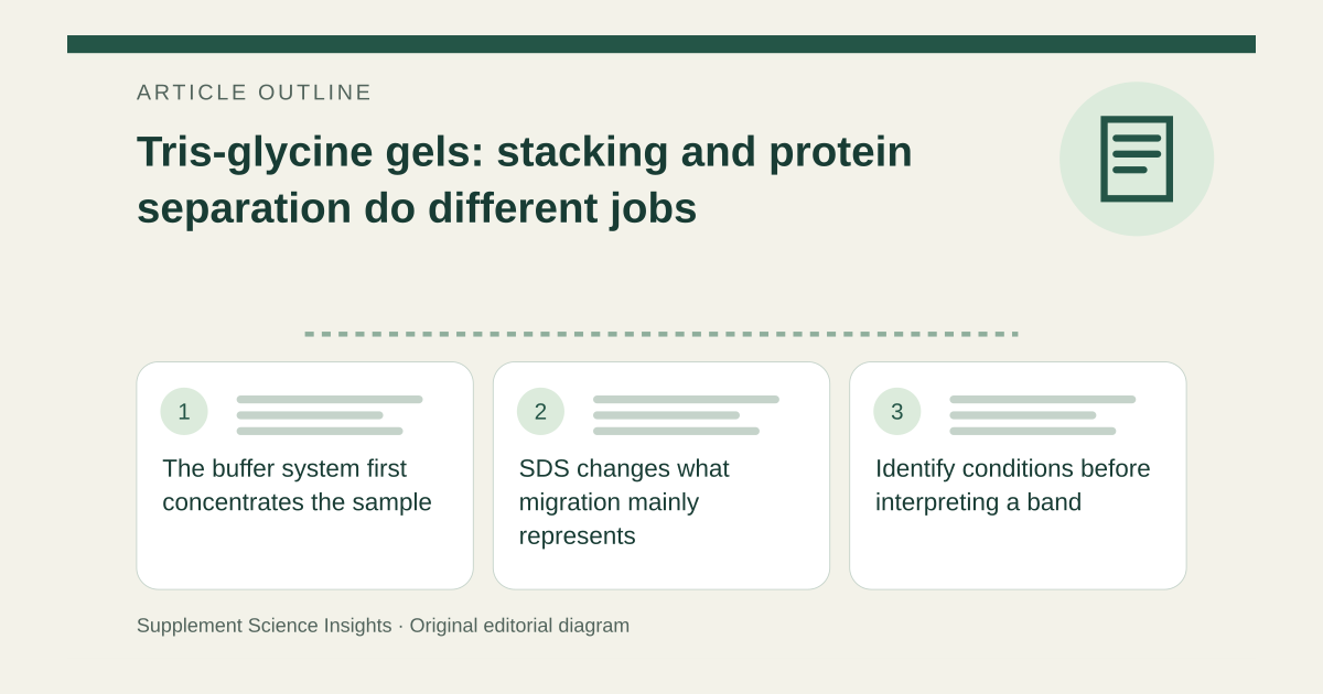
The buffer system first concentrates the sample
In the discontinuous system, chloride and glycine provide leading and trailing ion behavior that helps focus proteins into a narrow starting region. Tris supplies buffering and counterions. [1]
This stacking step improves the starting arrangement for separation. It is not itself a measurement of protein nutritional quality. [1]
SDS changes what migration mainly represents
SDS masks much of the proteins’ intrinsic charge and gives a broadly similar charge-to-mass relationship. Migration through the gel then depends primarily on size, allowing molecular-weight estimates against suitable standards. [1]
Without SDS, native protein charge and shape also matter. A band position cannot be interpreted using SDS-PAGE assumptions merely because the gel uses Tris and glycine. [1]
Identify conditions before interpreting a band
Imagine two proteins migrate differently in a native gel. That observation alone does not prove the faster protein has a lower molecular weight.
| Detail | Interpretation |
|---|---|
| Stacking | Concentrates the starting zone |
| SDS-PAGE | Mainly size-based separation |
| Native gel | Charge and shape also matter |
Sources for this article
- electrophoresis-biorad
Source consulted: 2026-09-18. This is a reference resource, not a claim that we conducted the studies or tested a product.