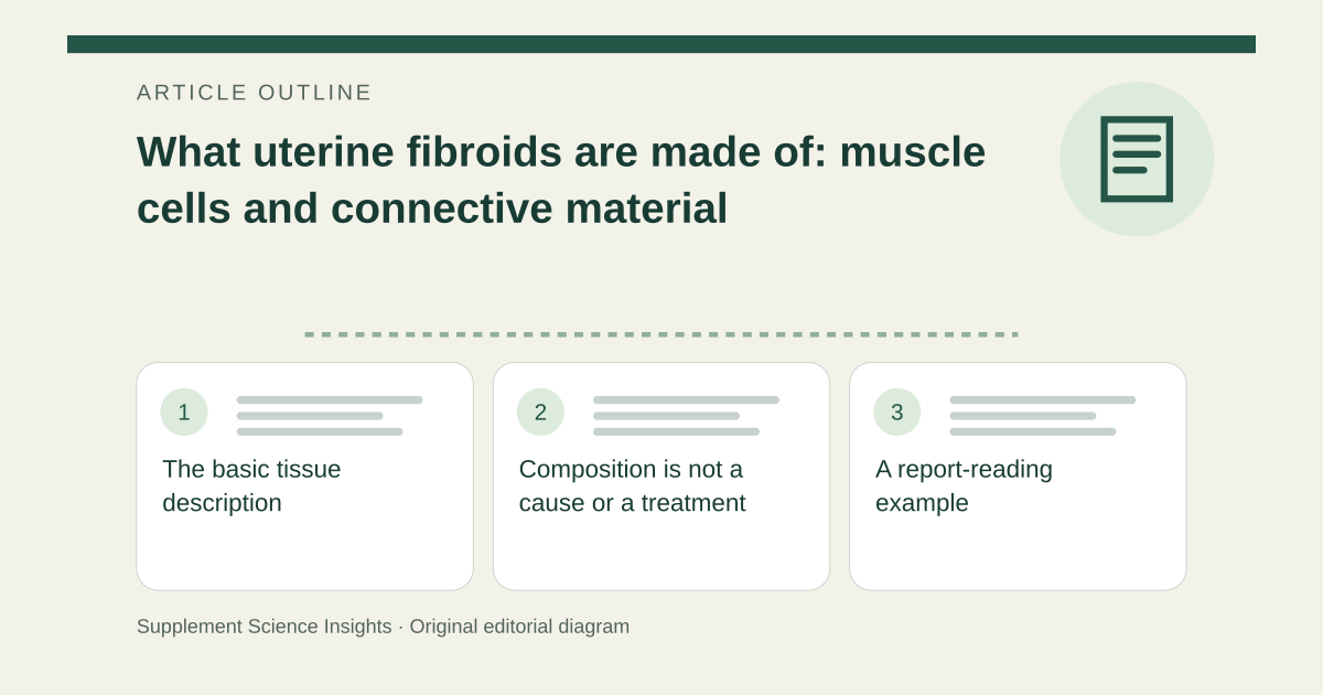
The basic tissue description
NICHD describes uterine fibroids, also called leiomyomas, as growths containing smooth muscle cells, fibroblasts and other material in or on the uterine wall. Fibroblasts are cells associated with production of extracellular matrix, including collagen-containing connective material. A fibroid is therefore an organized tissue growth rather than a capsule of one dietary ingredient. [1][2]
Composition is not a cause or a treatment
Finding a structural component in tissue does not establish that consuming that component created the growth. It also does not show that avoiding one protein or taking an enzyme dissolves it. NICHD describes multiple possible causal factors and diagnosis using clinical assessment and imaging; those questions go beyond naming the tissue constituents. [1]
A report-reading example
Imagine an ultrasound report identifies a uterine fibroid and the reader sees collagen mentioned in a separate explanation. That word does not identify the reason the fibroid developed or select a treatment. Symptoms, size, location and reproductive plans belong in a clinician-led discussion. Heavy bleeding or pain should not be managed by a self-selected collagen restriction alone. [1]
| Hypothetical information | What it establishes |
|---|---|
| Smooth muscle cells and matrix | Tissue composition |
| Collagen is present | Not proof of dietary causation |
| Imaging identifies a fibroid | Clinical context still needed |
Sources for this article
- Uterine Fibroids | NICHD - Eunice Kennedy Shriver National Institute of Child Health and Human Development
Source consulted: 2026-09-20. This is a reference resource, not a claim that we conducted the studies or tested a product. - The Extracellular Matrix of Animals - Molecular Biology of the Cell - NCBI Bookshelf
Source consulted: 2026-09-20. This is a reference resource, not a claim that we conducted the studies or tested a product.