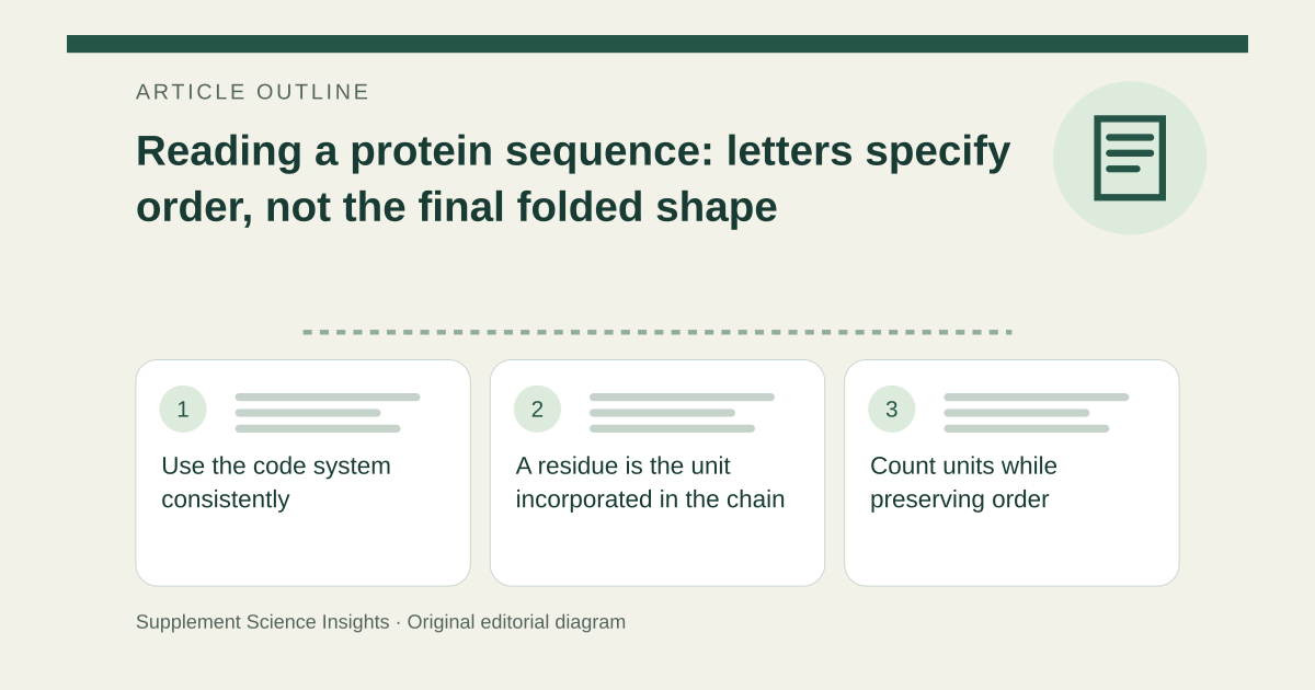
Use the code system consistently
Amino acids can be written with three-letter names such as Gly, Ala and Val, or one-letter codes G, A and V. The conventional sequence direction is from the N-terminus to the C-terminus. A letter is a code for a residue, not a dosage unit or an element symbol. [1][2]
For example, GAV corresponds to Gly–Ala–Val in that order. It contains the same residue inventory as VAG but has a different sequence. Order matters even when the count of each amino acid is unchanged. [1][2]
A residue is the unit incorporated in the chain
The term residue identifies an amino-acid unit as part of a peptide or protein. It should not be confused with leftover contamination. Peptide-bond formation changes the chemical context from that of an isolated free amino acid. [1][2]
A sequence describes primary structure. Folding, interactions among residues, assembly with other chains and chemical modifications provide additional levels of information. A short string of letters alone does not establish the whole protein’s function or a supplement’s clinical effect. [1][2]
Count units while preserving order
Imagine a fictional sequence Gly–Ala–Val. It has three residues and can be abbreviated GAV. Reversing it to Val–Ala–Gly still gives three residues, but it is a different ordered sequence rather than another spelling of the first.
| Feature | Interpretation |
|---|---|
| Gly–Ala–Val | Three-letter notation |
| GAV | One-letter notation |
| VAG | Different order |
Sources for this article
- OpenStax Biology 2e: Proteins
Source consulted: 2026-09-17. This is a reference resource, not a claim that we conducted the studies or tested a product. - OpenStax Organic Chemistry: Structures of Amino Acids
Source consulted: 2026-09-17. This is a reference resource, not a claim that we conducted the studies or tested a product.